Sponge Disease outbreak in Victoria
20th September 2020
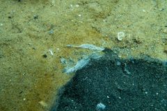
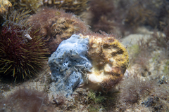
I originally thought it was a plastic shopping bag being dissolved by Bacteria, I flicked it with my fingers but only to see it smash to pieces with black dust inside resembling ash from a Coonara. The silver film resembled a thin piece of dissolved tissue by visual appearance. I didn't take much notice as I befevered the most likely possibility was local fishers discarding some type of burley or rubbish, I never thought to assume it to be a sponge.
This is the only infected specimen I have currently seen to date
12th October 2021
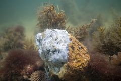
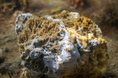
I came across it again not just a single specimen this time but probably ½ dozen of them. It appears to be some type of ‘Sponge Bacterial Disease’, although I’ve only seen it associate itself with one species of sponge it’s a possibility it may spread to more. This silver colour Bacteria completely covers the sponge eventually turning the sponge into black dust resembling ash from a fire place as mentioned above, then dislodges the sponge & gets carried by the currents to spread, well that's according to my current analysis based on this observation that was found almost washed to shore.


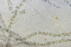
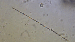
I took a sample of he infected tissue with the silver bacterial film while snapping some underwater photographs of the specimen then observed it under the microscope. I observed the chain bacteria through the microscope using a 100x oil objective with 10x eye piece (1000x Optical Magnification + cropping + Enlarging) & stacked the images from a short video with 600 frames. (The image quality still wasn't the best but I'll work on this)
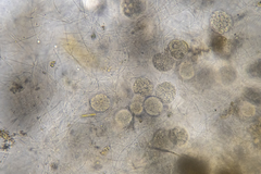
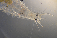
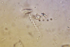
I also noted some Polychaeta (Top Right) that were thriving in the infected areas of the sponge.
After Posting to the Marine Research Group on Facebook I contacted Dr Richard Stafford-Bell who is the Principal Officer Invasive Marine Specie at Agriculture Victoria to make them aware of the outbreak, & Dr John Hooper who is international authority on sponges. John has been a great help and has helped shed a lot of light on the situation who has forwarded some emails & publications. One particular email reads...
Hello all,
Just saw the images in the link. One of my students, Gabriel Silva, is working with cultures of those fungi-like protist-thing from hell:
thraustochytrids (Stramenopiles). We believe they look very similar to what is seem in one of the diseased sponge samples (images attached).

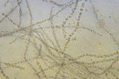
The filaments might be collagen filaments from an Ircinia, which can be the species in the picture (or Sarcotragus?). The large round structures are mature cysts from the traustochytrids, which can also be found as small isolated "cells". These "cells" can be quite small and produce large amounts of mucous substances, which can explain these filaments filled with small rounded structures.
They can be normally found associated with healthy sponges therefore we don't think that they are the disease, but a symptom. Due to some other factor (environmental change?) the sponges just lost the ability to keep it under control.
Best wishes, Marcio
Another email from the Porifera community reads...
Dear Colleagues,
I do not want to sound pessimistic. To solve the problem of the sponge disease is far from simple and it is too bad that it has negative impacts on the way of living of people. From my modest experience when studying a massive mortality in Mediterranean Ircinia spp, I can say that the decaying zones of the sponge body become secondarily colonized by a large variety of “secondary players” in the disease process. These organisms are different from the initial pathogen and they may include a variety of opportunistic bacteria and archaea, protists, fungi, etc. My impression is that the proliferation of these opportunistic secondary infections are more obvious then the initial pathogen. Thus, to elucidate the origin of the disease, it might be necessary to have access to individuals in their incipient stage of infection. Probably, an approach combining molecular biology and TEM will also be necessary for investigate those initial stages and prior to the manifestation of the general tissue decay. Even so, the task will not be easy. Please, just remember that we still do not know the etiological agents that caused in several waves the crash of the industry of sponge farming in the Mediterranean and also in the Caribbean over the XX century.
Best Wishes,
Manuel
20th October 2021
John helped me to get into contact with Lisa Goudie form the Melbourne Museum who I am planning to send specimens to (obtained by low impact research - No Permit Needed 'DEWLP')
It's easy to find the infected specimens in this particular area of Half Moon Bay as they appear to be numerous. My observation studies from this dive suggests that while some sponges may be recovering many more have been observed of containing this pathogen, I'm not sure if I'm just paying more attention or if some are newly infected - I'll keep an eye on it.
I collected samples from four separate specimens. I froze one and preserved a few for Lisa using 91% Methylated spirits (ethyl alcohol)with 9% purified water. Hopefully Lisa can identify the sponge, but the Bacteria probably needs to be alive to identify.
The microscopic findings from these samples suggests it's more complicated then I originally first thought, but what's got me worried is out of the three slides I observed I couldn't find any Thraustochytrids, perhaps this is because I wasn't observing the outermost layer? Or Perhaps the sample was mixed with the black powdery substance and what I observed is the decaying tissue of the sponge. Regardless of the sample I observed the species that I observed have me slightly worried, hence I hastily discarded the microscopic sample slides until I organize my workspace and collect more specimens.
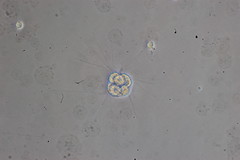
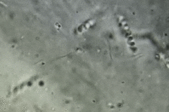
The Bacteria in images 1 and 2 are Spirochaetaes which can penetrate cell walls and are responsible for many nonvenereal diseases, these were in the millions and every space on the slide I observed these bacteria were present. Although I'm not sure what safety risks can be associated with viewing these bacteria's I will take safety precautions upon my next examinations of these organisms.
There were many other bacteria associated with the sponge as seen with the other images
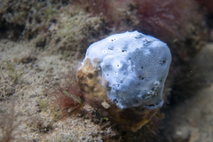
Other observed infected sponges include the below photos.
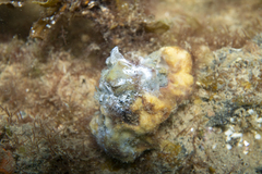
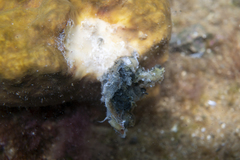
Again the Polychaeta worms are present, they love this kind of bacterial infected tissue. I have suspicions that they may be responsible for spreading the Pathogen....
To Be Continued


